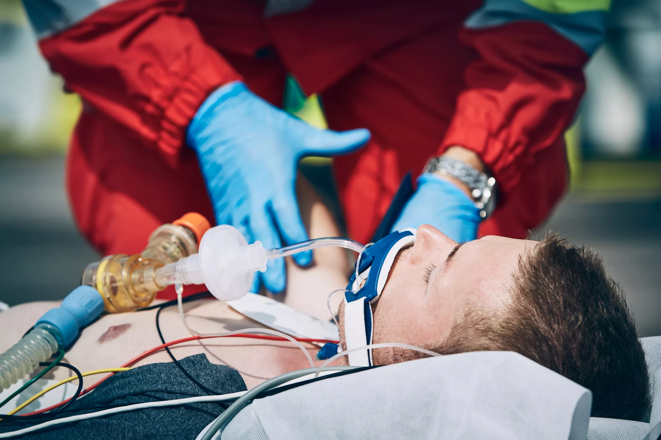
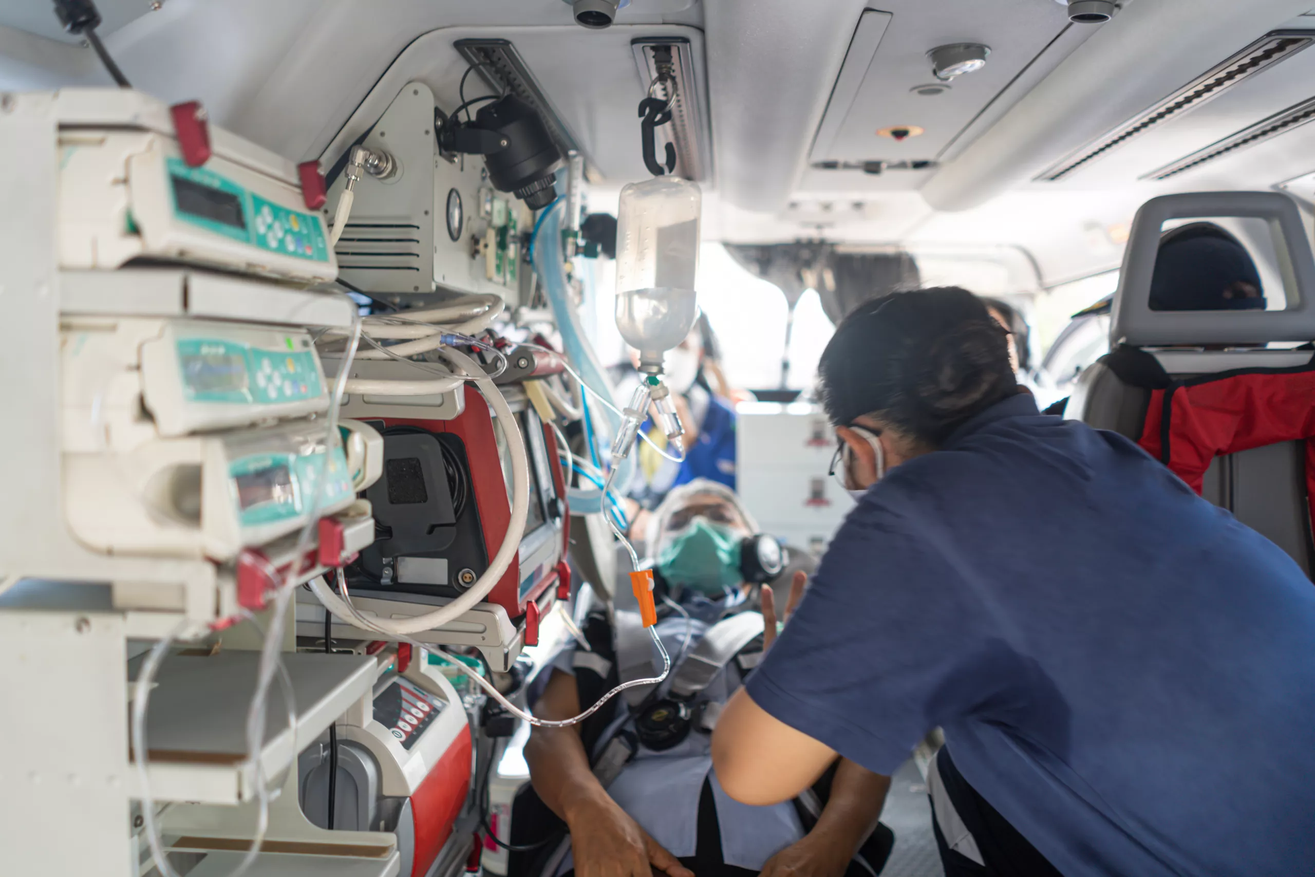
The role and consequences of mechanical ventilation during transport is something that must be both understood and respected. While it can free up much needed manpower and allow for more consistent delivery of oxygen and removal of carbon dioxide, it can also have negative effects if applied incorrectly. Broadly speaking, mechanical ventilation can be utilized to treat airway compromise and hypoventilation or apnea, but it also can be used to treat other conditions such as refractory hypoxia, hypercarbia or anticipated future airway compromise. When applied appropriately and in a timely fashion it can result in marked patient improvement. When applied inappropriately the consequences can be catastrophic.
Minute ventilation (MVe) is calculated using the formula: Ventilation x Tidal Volume. Normal minute ventilation at rest during normal spontaneous negative pressure breathing is somewhere between 4000 and 8000 ml/min. Under periods of physiological distress or compensation, this minute ventilation can double or triple. This is an important consideration during the application of mechanical ventilation. Not enough minute ventilation (think hypoventilation) can lead to too much carbon dioxide being retained and resulting in respiratory acidosis and a rightward shift on the oxyhemoglobin dissociation curve (OHDC). Too much minute ventilation (think hyperventilation) can result in too much carbon dioxide being removed and can result in respiratory alkalosis and a leftward shift in the OHDC.
The speed in which the mechanical breaths are delivered can also have a profound effect on the alveoli. Think about the bathroom door in your home: if you slam it repeatedly, eventually it will become damaged and fail to work properly. The same thing happens to our alveoli when we deliver too rapid or too forceful of a breath with a mechanical ventilator. This is called the Pendelluft effect. Say, for example there were 1000 people attending a movie that was scheduled to start at 7 pm. If they opened the theatre doors at 645, all 1000 people theoretically would rush in to be in their seats when the movie starts. Now let’s say the same movie is scheduled to start at 7 pm and the movie theatre allowed moviegoers to begin filling the theatre at 6 pm rather than 645. The same number and volume of attendees would seat themselves but they would enter at a slower pace and over a longer period of time. This is exactly what can happen to our alveoli, if we open them and shut them rapidly and forcefully they can become damaged and this can lead to alveolar collapse due absorptive atelectasis.
Another consideration during mechanical ventilation is dead space Dead space is defined as the overall surface area of the airway and lungs that do not actually participate in gas exchange. When breathing spontaneously without an airway device, ventilator circuit and all its attachments in place, this dead space involves the oropharynx, nasopharynx, hypo pharynx, larynx, trachea, right and left mainstem bronchi, the primary, secondary and some of the tertiary bronchi. These structures do not contain any alveoli and therefore cannot participate in gas exchange. These portions of the airway are called anatomical dead space. Physiological dead space is all the damaged, ineffective or collapsed alveoli that we see in conditions such as COPD, pneumonia, atelectasis etc. In mechanically ventilated patients, we have to add the ventilator circuit, the endotracheal tube and all attachments such as the flex connector, the exhalation filter and the ETC02 monitoring device to this dead space. All of these devices collectively are considered mechanical dead space. Because of all of this mechanical dead space we often see higher minute ventilation rates in mechanically ventilated patients than we see in spontaneously breathing patients. A minute ventilation rate of 100 ml/ kg of ideal body weight is often considered acceptable to account for the additional mechanical dead space in our intubated, mechanically ventilated patients. This assumes they have no additional minute ventilation requirements due to the body’s attempt to compensate for conditions such as illness or injury, metabolic acidosis, infectious conditions etc. Patients that are attempting to compensate for these types of issues may require minute ventilation calculations of 200-300 ml/kg of IBW to prevent further deterioration.
As you can see, placing a patient on a mechanical ventilator has many potential pitfalls and opportunities to cause further damage. Inappropriate ventilator settings can actually make our patients sicker rather than improving their status. It’s important to understand that providing the adequate rate of inhalation (I-time), minute ventilation (MVe), tidal volume (TV) can go a long way towards helping our patients rather than making them worse.
Think of it as the Goldilocks approach or theory: Not too much, not too little, but just right. An overall understanding of the advantages and pitfalls of applying mechanical ventilation principles to our mechanically ventilated patients can benefit them not only in the short term but also over their long term recovery by reducing downstream complications and improving morbidity and mortality.


