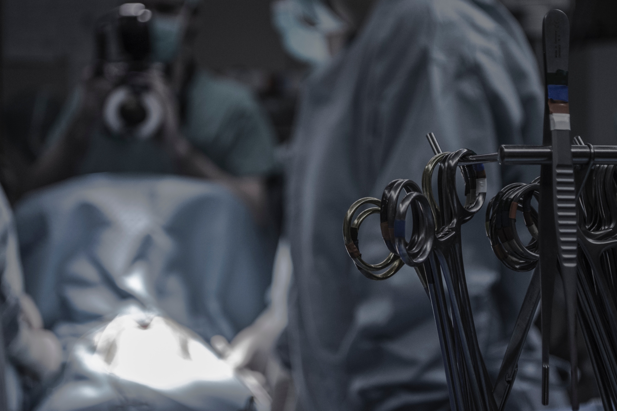
No longer will you ask your patient what kind of heart surgery they had upon seeing that sternotomy scar from an incision made before they saw through the length of your sternum and then reconnect it with wires after they’re done. If you haven’t already, you will have the patient that denies any heart problems and no scar is visible upon assessment. Your patient believes they don’t have a problem now because it has been fixed and it wasn’t surgery, it was interventional cardiology. Just like the patient that doesn’t have high blood pressure and then lists a dozen hypertension medications. An increasing number of cardiac procedures are being done under fluoroscopy (live imaging x-ray on a monitor) leaving a tiny scar where they accessed the vein or artery, usually femoral.
Cardiac catheterization was first performed when Werner Forssmann, in 1929, created an incision in one of his left antecubital veins and inserted a catheter into his venous system. He then guided the catheter by fluoroscopy into his right atrium. Subsequently, he walked up a flight of stairs to the radiology department and documented the procedure by having a chest x-ray performed. (Mueller & Sanborn, 1995)
Artificial pacemakers in 1932 were hand-wound, spring-driven generators that provided 6 minutes of pacemaking without rewinding. (Haddad, Houben, & Serdijin, 2006) Fully implantable pacemakers were available in 1958. The first implantable cardioverter defibrillator (AICD) came around 1980. All required a surgical incision usually in the left upper chest for the generator and a wire advanced via the left subclavian vein to the right atrium. Leadless pacemakers are inserted via the femoral vein and the entire unit, pacer and battery, is the size of a large vitamin pill and implanted in the right ventricle. (Mela & Singh, 2015) Someday you’ll be playing “Where’s Waldo” when you see pacer spikes on your EKG and no surgical scar or lump in the left upper chest.
Interventional cardiology can also replace heart valves, close an ASD (arterial septal defect) or VSD (ventricular septal defect). Atrial fibrillation might be treated with a Watchman device (Möbius-Winkler, et al., 2012)
A better assessment question might be “any tests or procedures on your heart?”
Bibliography
Haddad, S., Houben, R., & Serdijin, W. (2006). The evolution of pacemakers. IEEE Engineering in Medicine and Biology Magazine, 25(3), 38-48. Retrieved 5 20, 2023, from http://duteela.et.tudelft.nl/~wout/documents/emb2005.pdf
Mela, T., & Singh, J. P. (2015). Leadless pacemakers: leading us into the future? European Heart Journal, 36(37), 2520-2522. Retrieved 5 20, 2023, from https://ncbi.nlm.nih.gov/pubmed/26282468
Möbius-Winkler, S., Sandri, M., Mangner, N., Lurz, P., Dähnert, I., & Schuler, G. (2012). The WATCHMAN left atrial appendage closure device for atrial fibrillation. Journal of Visualized Experiments(60). Retrieved 5 20, 2023, from https://ncbi.nlm.nih.gov/pmc/articles/pmc3399494
Mueller, R. L., & Sanborn, T. A. (1995). The history of interventional cardiology: cardiac catheterization, angioplasty, and related interventions. American Heart Journal, 129(1), 146-172. Retrieved 5 20, 2023, from https://ncbi.nlm.nih.gov/pubmed/7817908
Micra TPS Pacer https://www.youtube.com/watch?v=RPrCDi-C_wU


