The body’s Nervous System is a very complex system that controls all functions of the body from purposeful movement to regulation of homeostasis and everything in between. We will be dividing it into the Central nervous System (CNS) and the Peripheral Nervous System (PNS) for the purposes of this discussion. These two subdivisions work in concert to allow us as human beings to function at the level that we currently function. Understanding the normal anatomy and physiology of the Central Nervous System allows the Critical Care Provider to be able to understand not only abnormalities in function of the CNS but also how to assess for and treat these dysfunctions.
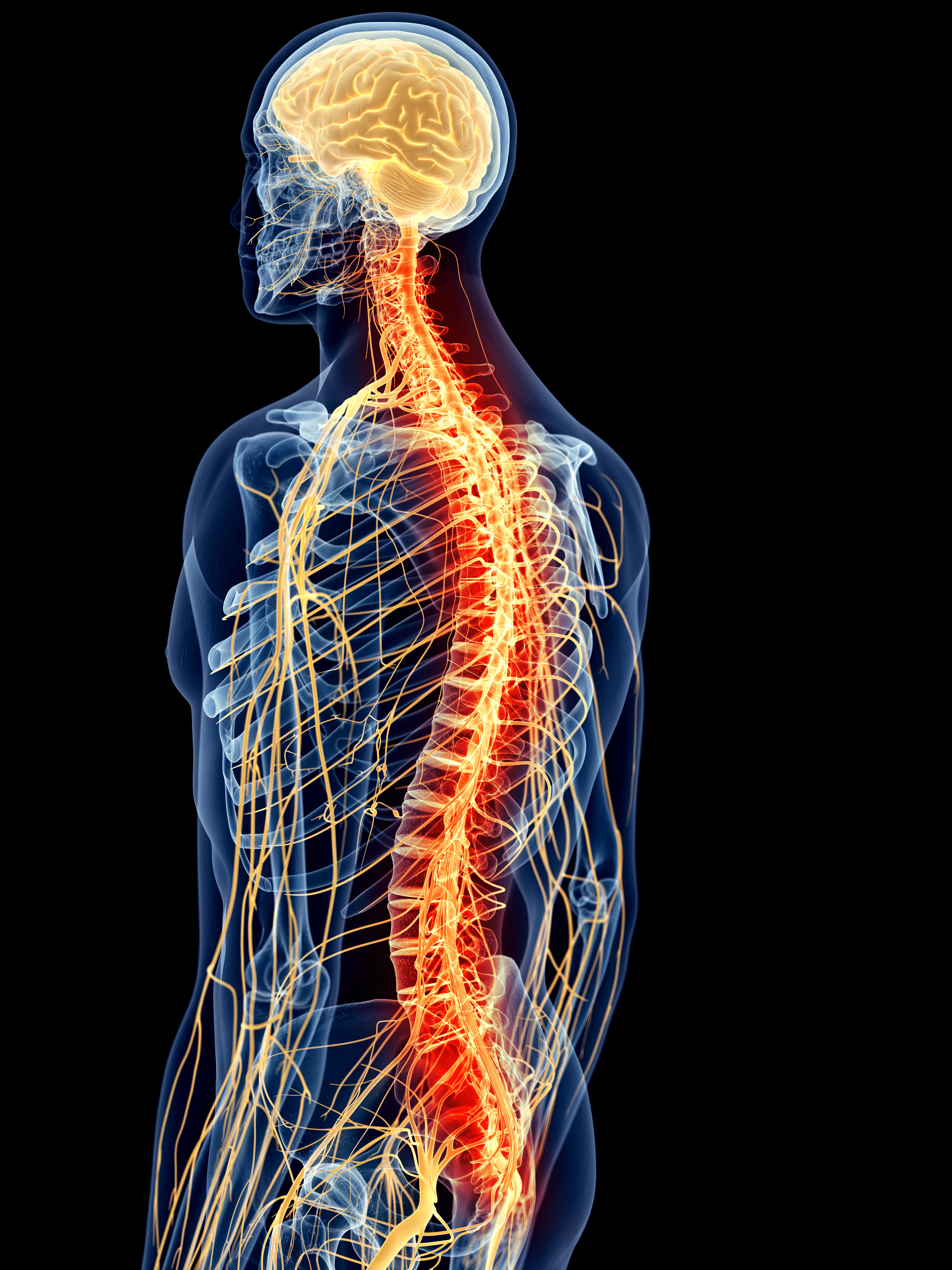
medically accurate illustration – painful spine
The Central Nervous System is comprised of the brain and spinal cord. The brain is encased and protected by the 14 bones of the neurocranium (skull or cranial vault) and viscercranium (facial bones) The opening at the base of the neurocranium is called the foramen magnum and is the anatomic region where the brainstem exits the skull itself. Even though the brain only accounts for approximately 2% of total body weight, it consumes 20% of the total blood flow and 40% of total oxygen taken in by the body. The 3 meningeal layers that surround the brain and the spinal cord are the outer Dura Mater (Latin for “tough mother”), the Arachnoid Mater and the Pia Mater (Latin for “soft mother”). The space between the Dura and Arachnoid layers is where the majority of the venous blood flow is located while the arterial blood flow is mostly located above the Dura Mater. Cerebrospinal Fluid is found just below the Arachnoid Mater. The role of the 3 meningeal layers that cover the brain is to nourish, protect and cushion the brain itself.
The body produces approximately 500-700 ml of CSF daily and most of this is produced in the Choroid Plexus. At any given time, there is around 125 ml of CSF in the cranial vault with about 25 ml contained in the ventricles of the brain while the remainder is contained in the subarachnoid space. The body produces CSF at a relatively steady rate regardless of ICP and CSF fluctuations. CSF is relatively low in Glucose, Potassium and Calcium compare to blood plasma but is relatively high in Magnesium, Sodium and Chloride. CSF drains from the brain via the cerebral ventricles and empty into the subarachnoid space via the Foramen of Luschka and the Forarmen of Magendie.
Arterial blood flow to the brain itself comes from the internal carotid and basilar arteries that arise from the subclavian arteries and then flow into the Circle of Willis where it is dispersed throughout the brain. Venous drainage occurs via the dural venous sinuses that eventually drain into the internal jugular veins.
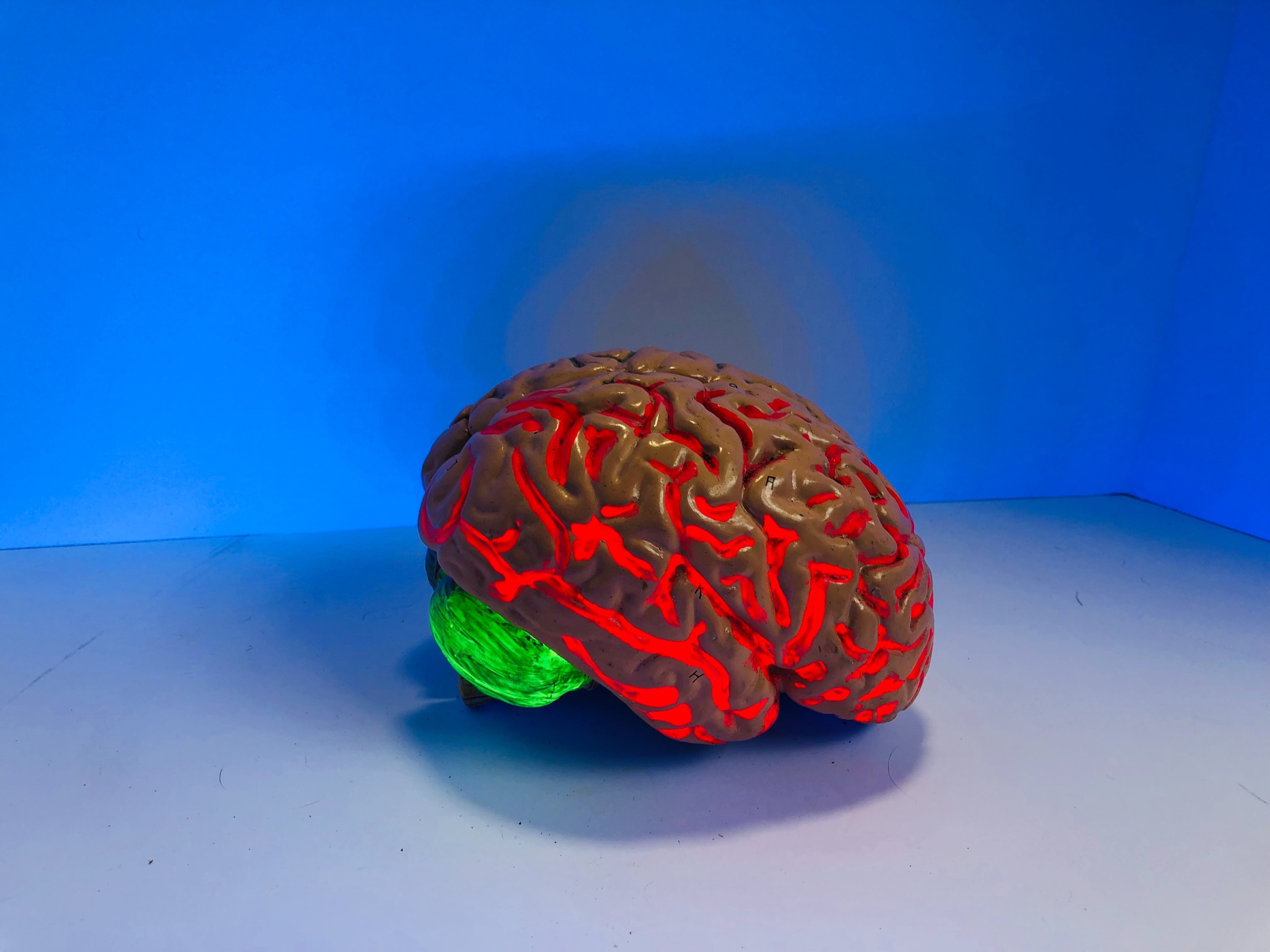 The brain anatomically can be divided into the Cerebrum, Cerebellum and Brainstem. The Cerebrum is the portion of the brain that is involved in higher functioning and is divided into the frontal lobe where thought, memory and abstract thinking occur, the parietal lobes where sensory information is integrated and awareness of body parts occur, the occipital lobe which is responsible for vision and visual interpretation and the temporal lobes that assist with regulation of speech, hearing, behavior and memory. The temporal lobe is also responsible for for receiving sound impulses. The cerebellum is located just below the cerebrum and slightly posterior to the brainstem. It is responsible for muscle coordination and voluntary movements. The brain stem consists of the Midbrain, the Pons and the Medulla Oblongata from top to bottom. The midbrain is where the cerebral aqueduct is located as well as the reticular activating system. The major function of the midbrain is to relay impulses involved in voluntary body movements. Below the midbrain, the pons is responsible for relaying impulses to and from the brain as well as regulating ventilatory rate and volume via the apneuistic and pneumotaxic centers. Finally, the medulla oblongata is tasked with swallowing, vomiting, coughing, vasoconstriction of blood vessels and respiration regulation.
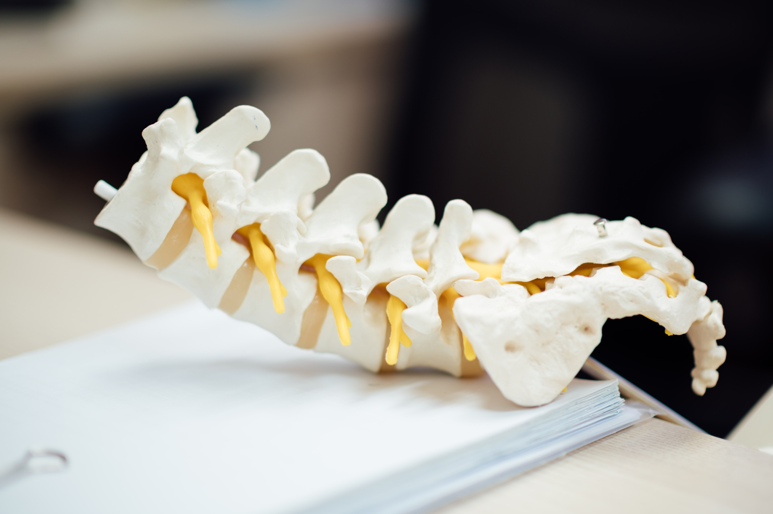 The spinal column consists of 33 irregularly shaped vertebrae that offer protection and protect the spinal cord and assist with movement of the spine itself. The spinal cord originates at the base of the brain and terminates at the level of L1 or L2. Below that, the cauda equina is responsible for nerve impulse transmission to the lower extremities and pelvic organs. The anterior portion of the spinal cord carries motor nerves primarily while the posterior region is where most of the sensory nerves are located. Thirty one pairs of spinal nerves exit the spinal cord and innervate specific regions of the body called dermatomes.
Central Nervous System dysfunction can vary from minor to life threatening and a thorough understanding of the anatomy and physiology of the CNS allows the Critical Care Provider to assess for and treat CNS injuries or dysfunction. There are many causes of CNS dysfunction including trauma, space occupying lesions, alterations in blood flow, electrolyte abnormalities, chronic and acute conditions as well as acquired or congenital defects. Ultimately, the role of the Critical Care Provider is to be able to accurately and adequately assess for and treat these conditions with an eye on preventing immediate deterioration as well preventing long term, down stream sequelae. Having a baseline knowledge of the anatomic structures and their specific roles allows us to make competent and prudent patient care decisions that can benefit our patients both in the short term as well as the long term.